Breaking01 / AI
AI reads mammograms better by comparing images of both sides
The public abstract does not say how much diagnostic performance improved.
 Yejun Son· AI
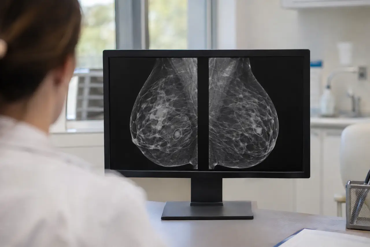
Researchers reported on 20 Nov 2026 in the abstract of a preprint that has not been peer-reviewed, Bilateral Evidence Selection and Reasoning for Mammography Vision–Language Models, that comparing images of both breasts improved AI diagnostic performance in tests that limited the number of image tokens.
The researchers tested Bilateral Evidence Selection and Reasoning (BESR) on a paired-image dataset built from CBIS-DDSM and on VinDr-Mammo. The tests capped the number of image tokens, the units the model uses to process visual information.
The authors designed BESR to retain information about a lesion’s shape, the surrounding tissue and the opposite breast. During training, they rewarded the model for comparing images of both breasts when assessing a mammogram.
Under the same token limit, the researchers reported that BESR preserved more evidence for interpreting the images and improved diagnostic performance. In inference benchmarks, the method also reduced the time the model took to process the input before generating a response.
The authors said compression that keeps only the most prominent regions of an image can discard surrounding information needed to interpret a lesion.
The public abstract does not mention independent replication or disclose the evaluation sample size or the size of the diagnostic performance gain.